Satellite Lab Course with Multiple Options for Attendance
- Live satellite format course with hands-on labs
- 20 contact hours, tuition $575
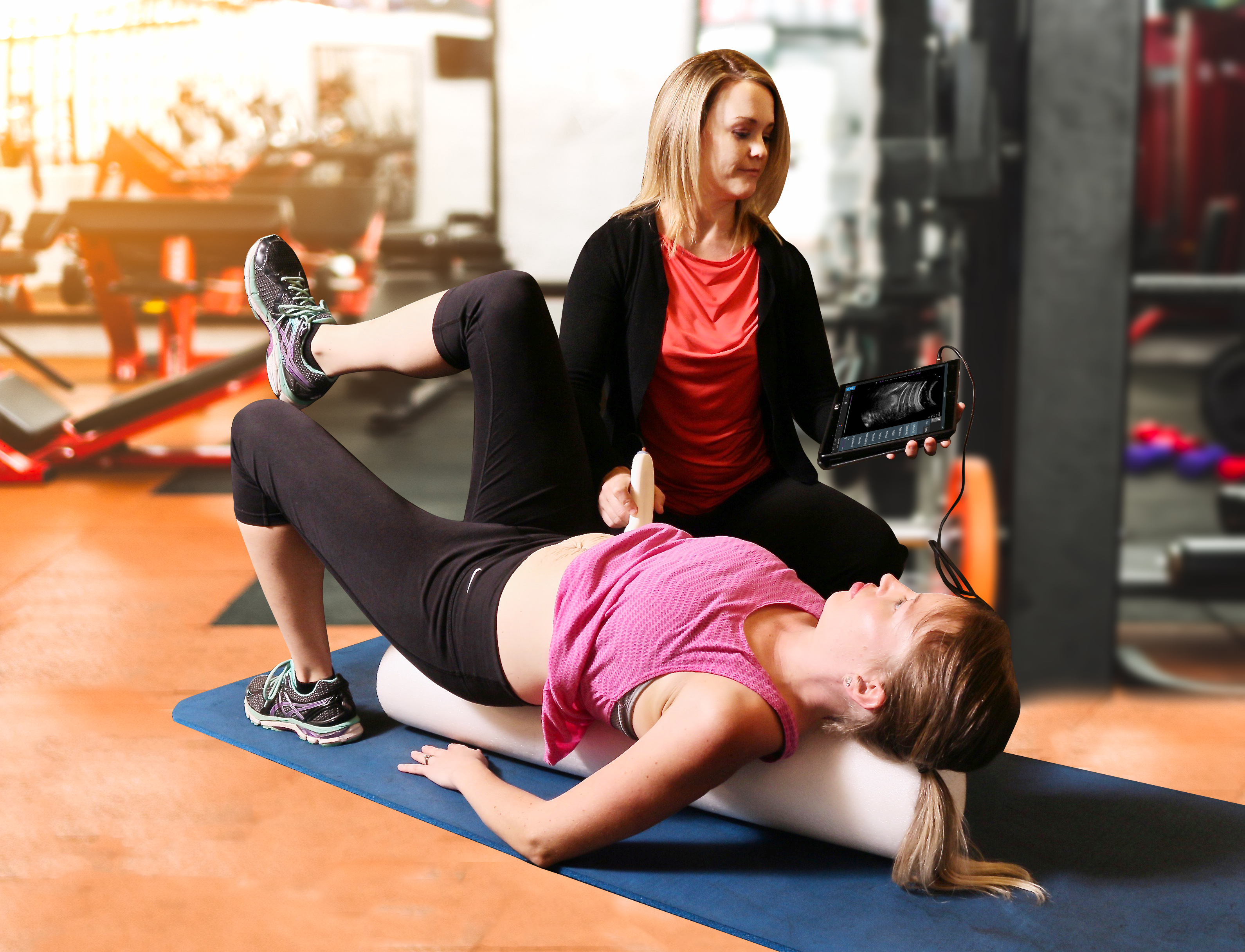
- Ultrasound equipment provided at all sponsored sites
- Competency-based educational model following based on international recommendations (Whittaker et al, 2019)
- Experienced faculty at all Host and Satellite locations
- Interested in hosting a Satellite, learning pod or serving as a teaching assistant? Contact us: [email protected]
- Prometheus Pathway RUSI users receive a 10% discount on all EchoGen Seminars. The discount code is located on the member's side of echogenportal.com
For more information, check out our Frequently Asked Questions page
Options for attending the course?
Host location: The instructor will be teaching in person from the host site and broadcasting with cameras via Zoom to all participating groups during the course. Learn more about being a Course Host
Satellite location: The lectures will be broadcast to the satellite facility with qualified teaching assistants in attendance. Learn more about being a Satellite Location
Self-hosted pod: Clinicians who have access to ultrasound equipment and a minimum of six months of imaging experience can create a learning pod of two or more participants at a private location. Clinicians who own equipment but lack the required experience need to have an approved TA to assist with the labs. See a list of Self-Hosting Requirements
If you have prior training and are Interested in being a TA for an upcoming course, email: [email protected]
Choose Your Preferred Site

- Ultrasound imaging allows for valid, reliable, efficient, and non-invasive measurement of motor control deficits of the deep stabilizing muscles that are associated with neuromusculoskeletal disorders.
- The ability to qualitatively assess motor control in real-time followed by visual feedback training has been shown to improve clinical decision-making and superior patient performance.
- We believe that the objective data obtained from ultrasound imaging will solidify the profession of physical therapy as the healthcare team's experts in musculoskeletal care.
About the Course
This two-day satellite-format course is designed as a competency-based model following international recommendations. Lectures followed by hands-on instruction for the utilization of rehabilitative ultrasound imaging (RUSI) for lumbopelvic dysfunction are the basis of the curriculum. The material will explore the scope of practice, ultrasound science, clinical evidence, regional anatomy, image generation, and ultrasound probe techniques for evaluating tissue morphology and motor pattern training. Clinical applications include a variety of lumbopelvic dysfunctions, including lumbosacral pain, urinary incontinence, diastasis rectus, pelvic organ prolapse, outlet dysfunction constipation, and chronic pelvic pain.
A prerequisite list of RUSI terminology and four hours of recorded lectures followed by a quiz must be completed prior to course onset. Saturday morning will begin with a live lecture on the use of ultrasound instrumentation immediately followed by technique demonstration and hands-on lab experience. Each clinical section will consist of technique validation, the clinical use of ultrasound imaging, interactive demonstration, hands-on practice and skills assessment. Faculty will be present during all lab sessions for assistance in learning. The afternoon of day two is specific to pelvic health diagnoses.
Topics to be covered:
- History of Ultrasound
-
Professional Considerations, ethics, and scope of practice
- Ultrasound 101: US physics, knobology, image generation, image interpretation
- Systematic techniques for performing an ultrasound scan on any region of the body
- Sonoanatomy and image interpretation
- M-Mode to perform objective testing
- Anterior abdominal wall
- Lateral abdominal wall
- Lumbar multifidus
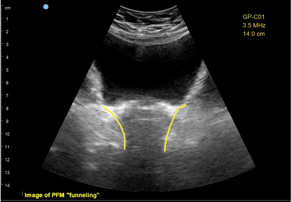
- Pelvic floor: Transabdominal approach
- Respiratory Diaphragm
- Pelvic floor: Transperineal approach for people with female anatomy
- Transperineal approach for the anterior and middle compartment of female anatomy
- Transperineal approach for people with male anatomy
- Transperineal approach for the posterior compartment of male and female anatomy
- Midline contents of the pelvis for prolapse, post-void residual, and megarectum
- Measurements and recording images of significant
- Using RUSI to market and grow your practice
Prerequisites:
Licensed health care provider with a solid grasp of lumbopelvic anatomy and rehabilitation of the muscles of local control. Prior ultrasound experience is not required. Pelvic floor training is required for participation in the transperineal labs. (Equipment required for self-hosted option).
Target Audience:
Course content is directed toward improving a clinician’s skill in the evaluation and treatment of lumbopelvic rehabilitation and is appropriate for all rehab clinicians. Physical Therapists, PT Assistants, Occupational Therapists, and Certified OT Assistants. Content is not intended for use outside the scope of the learner’s state license or regulations.
For more information, see our Frequently Asked Questions
Schedule By Time Zone
Eastern Standard Time
Saturday, Oct 12, 2024 - 10:00 am - 7:30 pm
Sunday, Oct 13, 2024 - 10:00 am - 6:30 pm
Central Standard Time
Saturday, Oct 12, 2024 - 9:00 am - 6:30 pm
Sunday, Oct 13, 2024 - 9:00 am - 5:30 pm
Mountain Standard Time
Saturday, Oct 12, 2024 - 8:00 am - 5:30 pm
Sunday, Oct 13, 2024 - 8:00 am - 4:30 pm
Pacific Standard Time
Saturday, Oct 12, 2024 - 7:00 am - 4:30 pm
Sunday, Oct 13, 2024 - 7:00 am - 3:30 pm

Course Equipment Sponsor
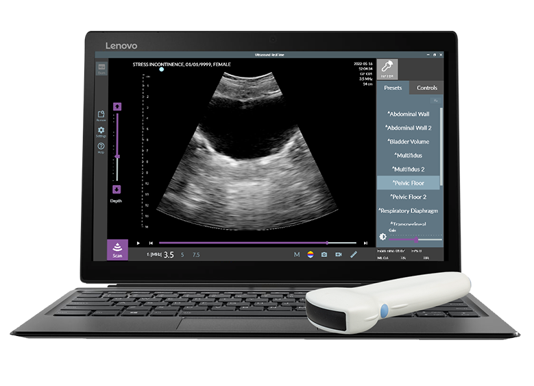
For information on the RUSI systems being used in this course, visit: The Prometheus Group
Example Curriculum
- Ultrasound Terminology
- RUSI Scope of Practice in Pelvic Health
- Slides for Pre-recorded Lectures
- Professional Considerations, Scope of practice and Safety (39:57)
- Science of Ultrasound Imaging (39:07)
- Performing an Ultrasound Scan (33:07)
- RUSI for the Rehabilitation Professional (25:46)
- Understanding Sonoanatomy (45:32)
- Prerequisite Material Quiz


